Part 1: Novel techniques and technology improve patient safety, care, and save health care dollars.
Groundbreaking computed tomography (CT) research by a group of Vancouver Coastal Health Research Institute (VCHRI) radiologists is sparking a significant radiology paradigm shift away from the 119-year old practice of x-ray imaging. The researchers are the first to publish findings in which new scanner technology and novel CT imaging techniques result in less radiation exposure to patients from a CT scan than from a conventional x-ray.
“The bar was always set at the radiation dose of the x-ray,” explains Dr. Hugue Ouellette, co-principal investigator and Chief Executive Officer of Vancouver Imaging.
“Once we were imaging lower than the dose of the x-ray, we essentially made a whole modality that has been used since 1895 obsolete.”
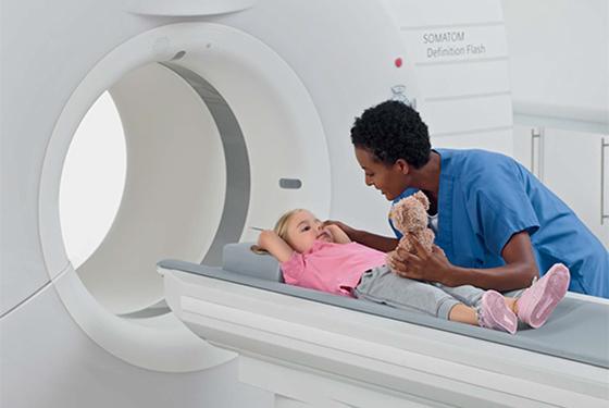
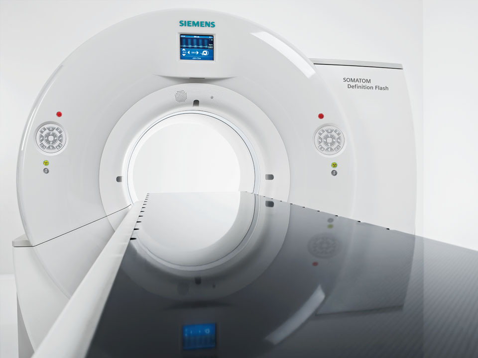
The group’s study*, “The Emergence of Ultra-Low Dose Computed Tomography and the Impending Obsolescence of the Plain Radiograph?”, was published in a recent issue of the Canadian Association of Radiologists Journal. The study details their use of the new Siemens CT scanner, “The Flash”. The scanner’s revolutionary technological capabilities come from its “Stellar Detector”, which transmits analog data with minimal wiring, making it possible to digitize the measured signals with virtually no interference. The team reverse engineered some aspects of modern CT technique by referencing how scans were performed five to ten years ago, with less complexity and fewer slices taken in each scan. This combination of cutting edge technology and novel technique consistently and dramatically lowers dose across many body parts.
“In terms of radiation exposure, the medical community previously learned that one CT scan equals approximately 300 to 400 chest x-rays,” explains Dr. Patrick McLaughlin, co-principal investigator and radiologist at Vancouver General Hospital. “There has been a genuine and sustained response to fears about radiation from CT scans, resulting in a huge collaborative effort by academic radiologists, community radiologists, and industry to immediately reduce doses to patients.”
The quest for lower CT scan doses also comes from their superiority as a diagnostic tool. X-rays, as a two-dimensional representation of a three-dimensional structure, do not provide as useful information to radiologists or referring physicians as CT scans.
“From an acute trauma surgical perspective, few trauma surgeons want to operate on a patient without seeing a CT scan first as it provides a roadmap of the critical injuries,” says Dr. Savvas Nicolaou, co-principal investigator, Chief Operating Officer of Vancouver Imaging, and head of emergency radiology at Vancouver General Hospital.
“The issue has always been radiation dose but if dose becomes a non-issue, you will want to order CT scans on the majority of patients in the acute setting rather than plain x-rays because it provides much more useful information required to save patients’ lives or perform the appropriate surgery.”
In addition to the critical role CT scans play in trauma situations, the team’s discovery will greatly benefit children and young women, who are more sensitive to the effects of the ionizing radiation, and patients requiring repeated scans.
Improved patient care equals cost savings
Adopting the higher technology required to produce the ultra-low dose CT scans will call for initial investment of health care dollars, however, patient care and overall health care system costs will ultimately be reduced.
“Coming to the emergency department and getting a CT scan rather than an x-ray increases confidence to either send someone to the operating room or back home appropriately,” says Dr. Ouellette.
“If patients are sent home incorrectly based on an x-ray, they may come back later due to complications, or, if they are sent to surgery incorrectly, there may be more health care dollars spent unnecessarily. Our new technology will improve diagnostic accuracy and move patients through the system faster, resulting in substantial savings.”
*Study co-principal investigators include Dr. Patrick D. McLaughlin, Dr. Hugue A. Ouellette, Dr. Luck J. Louis, Dr. Paul I. Mallinson, Dr. Timothy O’Connell, Dr. John R. Mayo, Dr. Peter L. Munk, and Dr. Savvas Nicolaou