
Main menu

  • About Us
    • Leadership
    • Our Team
    • Vision, Mission and Values
    • Health and Economic Impact
    • Research Impact Video
    • Strategic Plan
  • Our Research
    • Research Focus
      • Brain Health
      • Cancer
      • Digital Health and Artificial Intelligence
      • Heart Health
      • Healthy Aging and Mobility
      • Immune System
      • Injury and Rehabilitation
      • Lung Health
      • Mental Health and Substance Use
    • Research Centres and Programs
      • BC Centre on Substance Use
      • Centre for Aging SMART
      • Centre for Cardiovascular Innovation
      • Centre for Clinical Epidemiology and Evaluation
      • Centre for Lung Health
      • Djavad Mowafaghian Centre for Brain Health
      • Immunity and Infection Research Centre
      • International Collaboration On Repair Discoveries
      • M. H. Mohseni Institute of Urologic Sciences
      • Ovarian Cancer Research Centre
      • Community Research Program
      • Emergency Medicine Research Program
      • Hematology Research Program
      • Skin Research Program
      • Other Research Focus Areas
    • News and Stories
    • Researcher Directory
    • Events and Workshops
  • Research Services
    • New to VCHRI
      • Working at VCHRI
      • Regulations and Training
      • Membership with VCHRI
      • Learning and Development
    • Starting Your Project
      • Research Facilitation
      • Awards and Funding
      • Grant Management
      • Operational Approval
      • CST Cerner
    • Developing Your Project
      • Clinical Trials Administration
      • Clinical Research Unit
      • Research Privacy
      • Financial Policies and Procedures
    • Additional Support
      • Indigenous Health Research Unit
      • VCH-VCHRI AI Hub
      • Communications and Media Relations
      • Study Recruitment Support
      • Innovation and Industry Partnership
    • Internal Awards
    • Clinical Research
    • Indigenous Research
  • Participate in Research
    • Reasons to Participate
    • Participant Stories
    • Find a Study
    • Recruitment Support

User menu

  • Log in

Breadcrumb

  1. Home
  2. Our Research
  3. News and Stories
  4. Generating MRI images from ultrasounds scans

Generating MRI images from ultrasounds scans

Stories Sep 20, 2024 3 minutes

The novel application of artificial intelligence could address limited access to specialized scanning technologies and data for health research.

A study led by Vancouver Coastal Health Research Institute researcher Dr. Ilker Hacihaliloglu is the first to test the viability of a selection of image-to-image synthesizing technologies used to generate magnetic resonance imaging (MRI) images from ultrasound scans of prostate cancer. 

Dr. Ilker Hacihaliloglu is an associate professor in the Department of Radiology and Department of Medicine at the University of British Columbia, where he heads the Ultrasound Technology and AI for Healthcare (ULTRAi) research laboratory. He is also a researcher with the Djavad Mowafaghian Centre for Brain Health, Centre for Heart Lung Innovation, Centre for Aging SMART and the Institute for Computing, Information and Cognitive Systems.

Within the past two years, researchers have made incredible advances in image synthesizing technology. Similar to the popular OpenAI generative artificial intelligence (AI) algorithm that can create images and transform images into other images, synthetic image generation is a sophisticated AI computer algorithm. 

Researchers feed image synthesizing technology thousands of patient diagnostic scans to train it on identifying disease characteristics, such as images of variations of a certain type of cancer. The more images input into the algorithm, the better it will be at accurately generating a synthetic image, explains Hacihaliloglu. For example, the software image synthesizing technology examined in their study can quickly translate a single ultrasound scan into another medical diagnostic image, such as an MRI image, with the click of a button.

“Our goal is for image-to-image technology to advance to the point at which it can be used in health care settings to reduce wait-times and travel time for high-quality clinical imaging, such as MRI, and reduce radiation exposure from computed tomography (CT), X-ray or mammography scans,” states Hacihaliloglu. 

“This emerging technology has the potential to reduce the need for many MRI and CT diagnostic scans.”  

MRI technology uses magnetic field waves and computer-generated radio waves to take detailed images of internal tissue structures, including bones, blood vessels, organs and muscles. While non-invasive, the technology is presently only available as non-portable machines that may not be available outside of major city centres. The same is the case for CT scanners, which are a radiating technology that takes a series of detailed X-ray images of internal soft tissues and bones.

“Each image modality, whether it be X-rays, CT scans or ultrasounds, has certain advantages and disadvantages that could be necessary for clinicians to accurately diagnose and treat a patient.”

Ultrasounds, on the other hand, are a non-radiating technology that uses sound waves to produce images of tissues, such as organs, inside the body. Available as handheld, portable machines, ultrasounds are a convenient and accessible means of generating real-time pictures of internal tissues. Available in remote areas that may be far from other scanning technology, such as MRI or CT scanners, ultrasounds are an ideal choice as a rapid medium from which to generate synthetic MRI or CT images.

More research is needed to refine image-to-image software 

For their study, Hacihaliloglu and his team evaluated the quality of synthetic MRI images generated from 794 ultrasound scans of prostate cancer using five widely recognized image-to-image translation networks in medical imaging: 2D-Pix2Pix, 2D-CycleGAN, 3D-CycleGAN, 3D-UNET and 3D-AutoEncoder. Five doctors with over five years of experience reading MRI scans were then given a random selection of 15 synthesized MRI images. 

The following are examples of ultrasound and MRI scans as compared to synthetic MRI images generated from the corresponding ultrasound image above it.

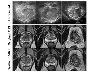
The team found that, while the synthetic MRI images were around 85 per cent similar to those of the MRI scans, they lacked too many important clinical features to be useful in diagnostics at this stage. 

“If the synthetic MRI images are accurate enough, we can one day use ultrasound scans to create MRI images.” 

“This technology has great potential to be used in clinical diagnostics, research and medication development,” says Hacihaliloglu. “As such, there is a pressing need for further refinement of image-to-image translation networks to enhance its performance.”
 

Researchers

Ilker Hacihaliloglu

Related Articles

A road map for early brain disease detection

Ask an expert: I know that X-rays and CT scans emit radiation. Should I be worried about getting multiple scans?

SPECT neuroimaging highly effective in helping diagnose PTSD and traumatic brain injury

Share:

  • Facebook
  • Linkedin
  • Twitter
  • Email

Related Research Centres/Programs

Centre for Aging SMART

Djavad Mowafaghian Centre for Brain Health

Get the latest research headlines in your inbox

Subscribe

Recent News and Stories

Type
Announcement

Celebrating the life and distinguished career of Dr. Marcel Dvorak

May 14, 2025
Type
Stories

More equitable representation needed in Parkinson’s research

May 9, 2025 parkinsons, patient engagement, women
Type
Stories

Gamified stroke recovery improves arm function

May 8, 2025 stroke, rehabilitation
See more news

Get updates!

Join our newsletter mailing list to stay up to date on features and releases.

Subscribe

Quick Links

  • News and Stories
  • Careers
  • Events
  • Media Enquiries

Follow Us

  • LinkedIn
  • X
  • YouTube

© 2025 VCHRI. All rights reserved.

  • Contact
  • Privacy Policy