Seasoned and up-and-coming scholars are at the forefront of technologies defining the digital wave of health research.
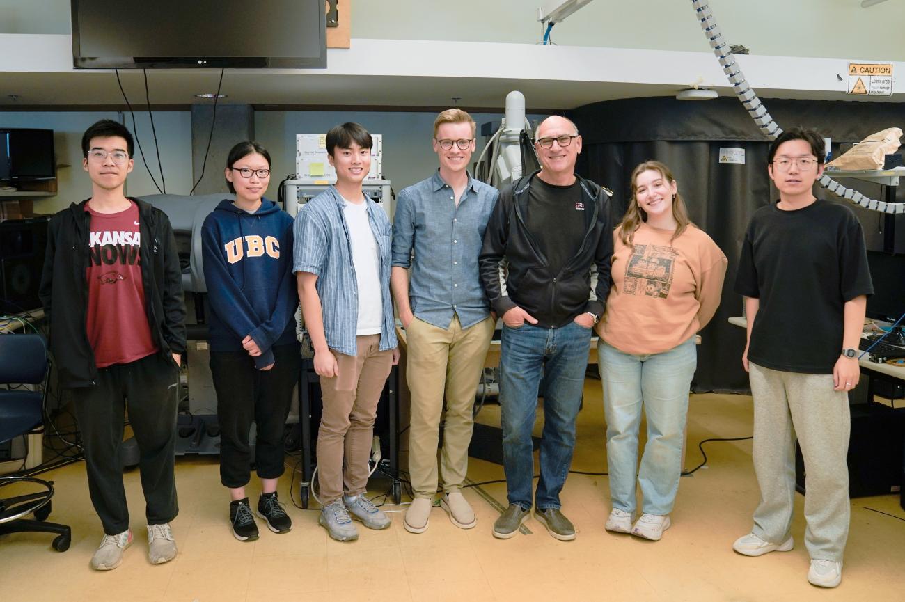
Walking into the Robotics and Control Laboratory (RCL), you first notice the rows of computers, many with researchers busily typing away in front of their screens. Beside desks and along the walls, robot arms and other apparatuses jut out at odd angles, some with wires cascading from them to racks of servers used to store dizzying amounts of data.
The RCL is a leading research facility in advanced machine-assisted medical applications. Headed by Vancouver Coastal Health Research Institute researchers Drs. Tim Salcudean and Purang Abolmaesumi, along with Institute for Computing, Information and Cognitive Systems director Dr. Robert Rohling, lab research focuses on medical robotics, telerobotics, ultrasound and image analysis. It is also a leading centre for image-guided diagnosis and interventions, with a goal to enhance precision medical procedures.
Technologies developed at the RCL have been integrated into products sold around the world. Applications have advanced real-time video imagery of the internal structures of the body for surgical and diagnostic procedures, such as in prostate cancer care.
Enhancing augmented and mixed reality care with the da Vinci surgical robot
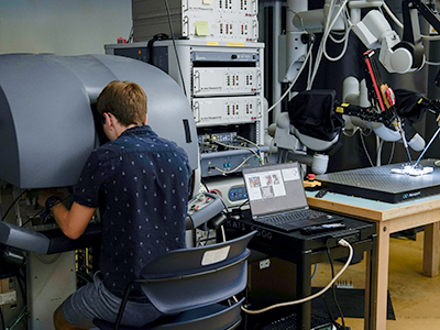
One of the advanced technologies investigated in the RCL is the da Vinci robotic surgical system. Using this robot, Wanwen Chen, a PhD candidate working in the lab, is making inroads in ultrasound applications in endoscopic surgery.
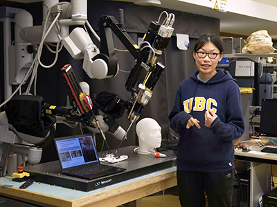
Chen’s software integration would make it possible to create a real-time, side-by-side view of the da Vinci’s camera video and ultrasound imagery. The point-of-care ultrasound (POCUS) transducer selected for this approach is a compact version of larger, traditional ultrasound machines, emitting sound waves to capture images of internal organs and tissues.
“The same technology that we developed for use with POCUS could produce side-by-side views of da Vinci robotic surgical system imagery with a computed tomography (CT) imaging scan or magnetic resonance imaging (MRI) scan,” notes Chen. “This advanced imaging integration offers additional layers of detail to surgeons, facilitating greater precision for enhanced patient outcomes and a reduced risk of removing excess tissue at the margins of a tumour or unintentionally coming into contact with vasculature, for example.”
Alexandre Banks, Master of Science candidate, is developing an AR hand-over-hand training system for the da Vinci technology. Banks records expert surgeons using the tool, then turns them into AR training videos. Trainees can see their own articulating arm tool at the same time as the digital overlay of the recorded one.
More accessible diagnostics with the novel application of ultrasound
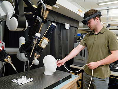
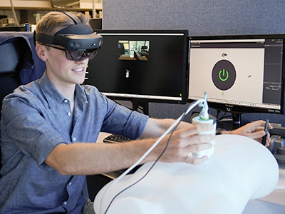
The tele-ultrasound prototype co-developed by David Black, a PhD candidate working in the RCL, and Salcudean, with master’s student Ryan Yeung and several undergraduate students, could enable expert sonographers or radiologists to guide non-experts located many kilometres away in performing an ultrasound scan. An MR headset and specially designed POCUS transducer would be used by a non-expert to conduct the scan under the remote guidance of an expert.
A live video feed from the non-expert’s headset-mounted camera would be fed to the expert via a video link. The expert’s haptic controller would guide the non-expert through a scan, with a digital overlay of the expert’s controller — in the form of a virtual transducer — appearing over the non-expert’s real-world environment.
“This force sensor that we developed measures changes in the magnetic field to measure transducer pressure exertion more accurately,” explains Black. “We are also investigating an option to replace the MR headset with a smartphone app to dramatically reduce the cost of the technology.”
Tara Kemper, a graduate student, is building on prior research into an elastography system that gauges organ stiffness through vibrations. Kemper’s novel approach uses ultrasound and a vibrating board placed below a patient to quantify liver fibrosis — scar tissue — and fat, with the hopes of creating a rapid non-invasive test to detect potentially deadly fatty liver disease.
“Different densities of tissues and liquids in the body produce different ripples, which we can then see in ultrasound images,” says Kemper. “The vibrations from the board, thus, enhance our ability to view tissue stiffness and potentially diagnose diseases, such as fatty liver disease, as well as others.”
Learn more about research underway at the Robotics and Control Laboratory.
"Behind the Lab Doors” is a Research Insider series that offers a behind-the-scenes glimpse into the labs of health researchers across VCHRI. These stories will explore research currently in the works, including ongoing and long-term studies that have the potential to directly impact quality of care for patients and clinicians. To feature a study from your VCHRI-affiliated lab, contact us at vchricommunications@vch.ca.