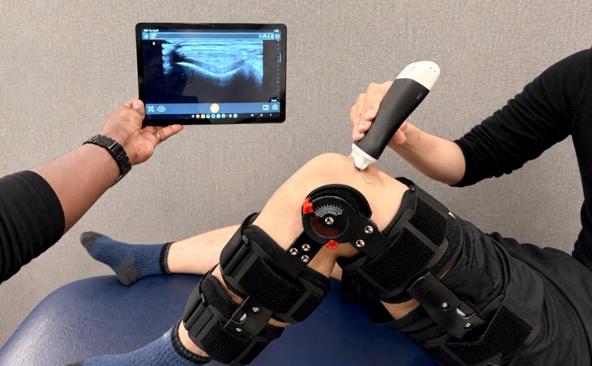
An AI-powered platform could give clinicians greater access to tools to prevent, monitor and treat knee osteoarthritis.
A new artificial intelligence (AI) tool spearheaded by Vancouver Coastal Health Research Institute researcher Dr. Ilker Hacihaliloglu could expand the early detection and treatment of knee osteoarthritis.
Hacihaliloglu and his team’s recent study introduced the MonoUNet AI tool, which enhances handheld ultrasound images and automatically highlights knee cartilage, making it easier for clinicians to see and measure changes associated with osteoarthritis. The technology could improve access to accurate diagnosis and ongoing monitoring in community clinics and rural settings, reducing the need for patients to travel to major urban centres.

Knee osteoarthritis affects more than 500 million people globally and around four million Canadians — approximately 14 per cent of the Canadian population. A progressive and incurable condition, knee osteoarthritis is caused by the wearing away of protective cartilage that cushions the knee joint.
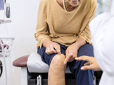
“Detecting changes in knee cartilage and beginning treatment as early as possible is imperative to preventing, slowing or stopping the progression of knee osteoarthritis.”
“MonoUNet could also be used to monitor changes in knee cartilage over time, helping doctors track how well new osteoarthritis treatments are working,” Hacihaliloglu adds.
The gold standard diagnostic tool for assessing changes in cartilage density is magnetic resonance imaging (MRI). However, MRI machines are in high-demand, often have long wait times and are only available in large urban centres. As a result, more portable and affordable options are a high priority for optimal knee osteoarthritis diagnostics, treatment monitoring and prevention.
Expanding access to knee imaging with handheld ultrasound
A promising complement to MRI scans for knee imaging is point-of-care ultrasound (POCUS). POCUS’s lightweight system includes a wireless, handheld transducer that emits sound waves to capture images of internal structures in the body. While similar in many ways to a standard ultrasound machine, POCUS is more portable, affordable and able to relay images to electronic devices like a tablets, smartphones or laptops.
However, even small differences in how the handheld POCUS system’s transducer is positioned can significantly affect image quality, making it more difficult to obtain clear and consistent images.
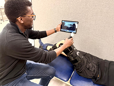
Hacihaliloglu’s MonoUNet machine learning software was designed to take images collected using POCUS a step further. Running on a tablet, laptop, smartphone or in the cloud, the software provides real-time guidance while a scan is being performed, helping users capture clear, consistent images.
Once a scan is complete, the AI automatically enhances the images, identifies and outlines the knee cartilage and provides measurements that support clinicians in assessing joint health and tracking changes over time.
“Rather than relying only on the brightness of an ultrasound image, MonoUNet looks for underlying structural patterns that remain consistent even when image quality or scanning conditions change,” Hacihaliloglu explains.
“Similar to putting on night-vision goggles, MonoUNet’s machine learning capabilities enhance POCUS images, making subtle features and tissue boundaries easier to see and interpret.”
“The enhanced images from MonoUNet help clinicians obtain clearer, more consistent images and make more confident assessments,” adds Hacihaliloglu.
A high-performing technology that outmatches its peers
In their study, researchers evaluated MonoUNet using ultrasound images collected at multiple sites with four different ultrasound devices, including POCUS systems. The model’s design was up to 700 times more efficient than comparable models, requiring as much as 2,000 times less computational power while maintaining a degree of accuracy comparable to medical imaging experts.
“Because MonoUNet can run on an electronic device connected to a portable ultrasound, it could bring high-quality imaging for knee cartilage analysis to patients in community settings and in rural areas.”
One of MonoUNet’s biggest advantages was its ability to work across different ultrasound systems. AI models are often trained using images from one machine and perform less accurately when used with another. By emphasizing structural information in images, such as the edges and borders around cartilage, MonoUNet maintained a high degree of performance even when tested on images collected with different devices under varying scanning conditions.
“MonoUNet’s ability to provide rapid results could also lead to real-time feedback during a POCUS scan, relaying information to the transducer operator on how much pressure to apply to capture images of the highest possible quality,” Hacihaliloglu emphasizes.
The research team is already building on their findings by developing even more compact AI architectures and exploring possible ways to combine them with MonoUNet’s structural image analysis. They also plan to validate the technology in larger and more diverse patient populations and investigate additional imaging features that could further improve performance.