
Main menu

  • About Us
    • Leadership
    • Our Team
    • Vision, Mission and Values
    • Health and Economic Impact
    • Strategic Plan
  • Our Research
    • Research Focus
      • Brain Health
      • Cancer
      • Cardiovascular Conditions and Diseases
      • Digital Health
      • Health Policy
      • Immunity and Infectious Diseases
      • Joint Health and Mobility
      • Respiratory and Lung Health
      • Spinal Cord Injury and Rehabilitation
    • Research Centres and Programs
      • BC Centre on Substance Use
      • Centre for Aging SMART
      • Centre for Cardiovascular Innovation
      • Centre for Clinical Epidemiology and Evaluation
      • Centre for Lung Health
      • Djavad Mowafaghian Centre for Brain Health
      • Immunity and Infection Research Centre
      • International Collaboration On Repair Discoveries
      • Ovarian Cancer Research Centre
      • Vancouver Prostate Centre
      • Community Research Program
      • Emergency Medicine Research Program
      • Hematology Research Program
      • Skin Research Program
      • Other Research Focus Areas
    • News and Stories
    • Researcher Directory
    • Events and Workshops
  • Research Services
    • New to VCHRI
      • Working at VCHRI
      • Regulations and Training
      • Membership with VCHRI
    • Starting Your Project
      • Research Facilitation
      • Awards and Funding
      • Grant Management
      • Operational Approval
      • CST Cerner
    • Developing Your Project
      • Clinical Trials Administration
      • Clinical Research Unit
      • Research Privacy
      • Financial Policies and Procedures
    • Additional Support
      • Education and Training
      • Communications and Branding
      • Media Relations
      • Study Recruitment Support
      • Innovation and Industry Partnership
    • Internal Awards
    • Clinical Research
    • Innovation and Partnership
  • Participate in Research
    • Reasons to Participate
    • Participant Stories
    • Find a Study
    • Recruitment Support

User menu

  • Log in

Breadcrumb

  1. Home
  2. Our Research
  3. News and Stories
  4. Machine learning takes disease detection to the next level

Machine learning takes disease detection to the next level

Stories Jun 21, 2018 4 minutes

Computers enhance traditional diagnosis, but more research is needed.

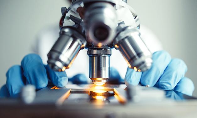
Inside the Department of Pathology and Laboratory Medicine at Vancouver General Hospital (VGH), a clinician peers through a microscope. She is looking at a thin slice from a block of paraffin wax embedded with tissue samples, searching for clues to cure disease. VGH neuropathologist and VCHRI researcher Dr. Stephen Yip calls this the analogue version of histology—the study of the structure of tissues at the microscopic level. Research he and colleagues are conducting is paving the way for histology 2.0 in the age of digital technology.

Yip and Adrian Levine, a resident of pathology and laboratory medicine at VGH, are researching how software programs that use deep learning can add a layer of quality control to diagnosing diseases from tissue samples. Deep learning here refers to computer software that uses algorithms to recognize what data represents, such as curves and lines in an image. This is similar to technology found in self-driving cars, which uses machine learning to detect features in the landscape. 

“The advantage of the software we are using is that it might see subtle features that our human eyes miss,” says Yip, medical director of the Clinical Genetics and Genomics Laboratory at BC Cancer.

The deep learning tools Yip and Levine are using have been around for a decade, but only in the past few years have they become popular for diagnosing diseases, such as cancer, from tissue samples. Advances in an area of deep learning called convolutional neural networks (CNNs) have been pivotal to this newfound application. 

CNNs are computer algorithms that identify specific characteristics in images using a layering technique. This makes CNNs ideal for medical specialties that rely heavily on image-based data—such as radiology and pathology—for interpreting information and making diagnoses. They enable computers to accurately differentiate between, for example, images of cancerous and non-cancerous cells. 

Research is fueling the new frontier of disease identification

To make it possible for computers to identify disease, millions of images of tissue samples need to be uploaded as digital copies. These images train the deep learning software to identify different medical conditions based on previous diagnoses. 

“The gold standard will be to make sure the software works on new real-world tissue samples.” 

While all the images in the database provide essential baseline information, the long-term hope is for this technology to be able to identify diseases from images it has never seen before. 

At some centres, radiologists already apply machine learning as part of their day-to-day work, including for examining X-rays and CT scans. By comparison, histologists have been peering through microscopes at tissue samples sandwiched between panes of glass since the mid-1800s, which Yip calls the “analog” way of doing things in the new digital frontier.

“We still have to digitize a huge number of slides to provide the training data our software needs to reach the gold standard of disease identification.”

Research, including a paper submitted by Levine, Yip and colleagues called “Machine learning in pathology: a primer on techniques and applications,” discusses examples where combining CNNs with a pathologist’s diagnosis improved the accuracy of the diagnosis by several percentage points. In one example, the CNN was able to more accurately distinguish between different types of lung cancer than its human pathologist counterpart.

The ability of machine learning to aid in cancer diagnosis was the topic of a news and views piece by Yip published in the journal Nature on March 14, 2018.

CNN technology is still in the early development stages as a clinical diagnostic tool. Scientists do not yet know with 100 per cent certainty whether there may be inherent biases in its programming that could interfere with a proper diagnosis. In other words, at least for the time being, diagnoses will still need to be signed off by a qualified human.

“It is very difficult to teach computers how to recognize images of diseases," says Levine. "A typical picture contains millions of pixels, which computers need to convert into data sets to interpret. Rigorous trials are still needed before CNNs are more widely used.”


Researchers

Stephen Yip

Related Articles

Tele-robots for care delivery at a distance

Computer-aided detection and diagnosis of colorectal cancer

Wearable technology for respiratory disease monitoring

Share:

  • Facebook
  • Linkedin
  • X
  • Email

Get the latest research headlines in your inbox

Subscribe

Recent News and Stories

Type
Stories

Leaps forward are still needed to improve Indigenous health care experiences

Sep 27, 2023 indigenous health
Type
Stories

Cancer driver discovery opens up treatment pathways for rare pediatric disease

Sep 25, 2023 cancer, treatment options, children
Type
Stories

People in profile: Karina Thiessen

Sep 20, 2023 people feature
See more news

Get updates!

Join our newsletter mailing list to stay up to date on features and releases.

Subscribe

Quick Links

  • News and Stories
  • Careers
  • Events
  • Media Enquiries

Follow Us

  • X
  • LinkedIn

© 2023 VCHRI. All rights reserved.

  • Contact
  • Privacy Policy