
Main menu

  • About Us
    • Leadership
    • Our Team
    • Vision, Mission and Values
    • Health and Economic Impact
    • Research Impact Video
    • Strategic Plan
  • Our Research
    • Research Focus
      • Brain Health
      • Cancer
      • Digital Health and Artificial Intelligence
      • Heart Health
      • Healthy Aging and Mobility
      • Immune System
      • Injury and Rehabilitation
      • Lung Health
      • Mental Health and Substance Use
    • Research Centres and Programs
      • BC Centre on Substance Use
      • Centre for Aging SMART
      • Centre for Cardiovascular Innovation
      • Centre for Clinical Epidemiology and Evaluation
      • Centre for Lung Health
      • Djavad Mowafaghian Centre for Brain Health
      • Immunity and Infection Research Centre
      • International Collaboration On Repair Discoveries
      • M. H. Mohseni Institute of Urologic Sciences
      • Ovarian Cancer Research Centre
      • Community Research Program
      • Emergency Medicine Research Program
      • Hematology Research Program
      • Skin Research Program
      • Other Research Focus Areas
    • News and Stories
    • Researcher Directory
    • Events and Workshops
  • Research Services
    • New to VCHRI
      • Working at VCHRI
      • Regulations and Training
      • Membership with VCHRI
      • Learning and Development
    • Starting Your Project
      • Research Facilitation
      • Awards and Funding
      • Grant Management
      • Operational Approval
      • CST Cerner
    • Developing Your Project
      • Clinical Trials Administration
      • Clinical Research Unit
      • Research Privacy
      • Financial Policies and Procedures
    • Additional Support
      • Indigenous Health Research Unit
      • VCH-VCHRI AI Hub
      • Communications and Media Relations
      • Study Recruitment Support
      • Innovation and Industry Partnership
    • Internal Awards
    • Clinical Research
    • Indigenous Research
  • Participate in Research
    • Reasons to Participate
    • Participant Stories
    • Find a Study
    • Recruitment Support

User menu

  • Log in

Breadcrumb

  1. Home
  2. Our Research
  3. News and Stories
  4. Novel AI model explains retinal sex difference

Novel AI model explains retinal sex difference

Stories Mar 20, 2024 3 minutes

The approach could lead to further biomarker discoveries to assist in disease diagnostics and early treatment interventions.

Artificial intelligence (AI) applications are revolutionizing health diagnostics and precision medicine, yet how they identify patterns of disease in layers of data has largely remained a mystery. Research led by Vancouver Coastal Health Research Institute researcher Dr. Ipek Oruç revealed for the first time AI behaviours that differentiated between female and male retinal images, paving the way for other novel biomarkers of disease to aid in precision care. 

Dr. Ipek Oruc is an associate professor in the Department of Ophthalmology and Visual Sciences; associate member of the School of Biomedical Engineering; investigator with the Data Science Institute; and director of the NOVA Lab at the University of British Columbia. She is also a principal investigator with ICORD.

An expert in visual neuroscience and artificial intelligence applications of retinal image analysis, Oruç’s PNAS Nexus study charts a path for other research teams to peek under the hood of AI algorithms to understand how the algorithm was developed in order to improve patient diagnostics and outcomes using medical imaging.

“Our research opens the black box of AI, setting the stage for future research to apply this methodology to leverage its potential and identify previously unseen characteristics of various conditions.”

Oruç and her team’s proof-of-concept study examined a convolutional neural network (CNN) model trained to classify patient sex in retinal images. CNNs are a type of deep neural network AI that can mimic human cognition in image processing and classification at superhuman speeds, processing thousands of images in a matter of seconds. The technology is already applied to a number of different AI algorithms using medical imaging for the detection and classification of conditions such as cancers and heart disease.

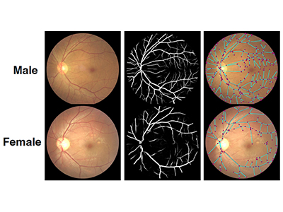
The CNN used in Oruç’s study was trained to detect male and female retinal scans taken using a specialized fundus camera, a mainstay tool of optometry and ophthalmology used to capture images of the retina and other eye features. 

Ophthalmologists and nonexperts better able to detect retinal sex difference

The research team developed and applied a novel methodology to discover for the first time retinal features that differentiate between male and female eyes. They tested 14 exploratory research questions that were derived from the behaviours and decisions of the CNN in what investigators termed the “Inspiration” phase of the model. Nine of the hypotheses revealed significant findings, five of which were verified.

These biomarkers of sex difference in retinal images included greater retinal vascularization and a darker ring around the optic disk region in male retinas as compared to female retinas.

These two images show subtle differences between male and female retinal images. The male image has a darker area around the optic disk, which appears as a bright spot in the images here, as well as more vasculature and nodes.

Researchers then shared these distinguishing features with 26 expert ophthalmologists and 31 nonexperts. Prior to receiving this information, both groups’ ability to detect sex difference in retinal images averaged 50 per cent. After receiving training on the distinguishing characteristics between female and male retinal images discovered through Oruç’s novel methodology, both groups were able to identify the sex of the retinal images with approximately 66 per cent accuracy in the post-training block. This significant improvement is still a ways away from a 100 per cent detection rate, which indicates that additional differences have yet to be discovered to enable greater accuracy, Oruç says.

"Our findings showcase an opportunity for biomarker discovery through CNN applications, with the added benefit of equipping medical practitioners with new diagnostic options that can be added to their clinical toolkit.”

“We are now investigating other biomarkers present in fundus images that can be identified from CNNs, such as whether changes in the eye might signal a risk of stroke or dementia.”

Oruç is also currently working on a study to apply this novel methodology to identify biomarkers in women that could lead to improvements in disease detection and treatment.
 

Researchers

Ipek Oruc

Related Articles

AI innovation puts VCHRI scientist at the forefront of global drug discovery avenues

Generating MRI images from ultrasounds scans

Machine learning for handheld cardiac ultrasounds

Share:

  • Facebook
  • Linkedin
  • Twitter
  • Email

Related Research Centres/Programs

International Collaboration on Repair Discoveries

Get the latest research headlines in your inbox

Subscribe

Recent News and Stories

Type
Announcement

Celebrating the life and distinguished career of Dr. Marcel Dvorak

May 14, 2025
Type
Stories

More equitable representation needed in Parkinson’s research

May 9, 2025 parkinsons, patient engagement, women
Type
Stories

Gamified stroke recovery improves arm function

May 8, 2025 stroke, rehabilitation
See more news

Get updates!

Join our newsletter mailing list to stay up to date on features and releases.

Subscribe

Quick Links

  • News and Stories
  • Careers
  • Events
  • Media Enquiries

Follow Us

  • LinkedIn
  • X
  • YouTube

© 2025 VCHRI. All rights reserved.

  • Contact
  • Privacy Policy